

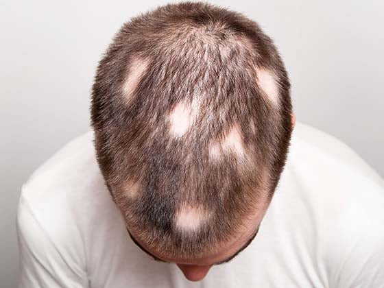


- What Is Epidermodysplasia Verruciformis?
- How EV forms?
- Tree Man Syndrome
- Common EV lesions
- Clinical diagnosis steps
- EV treatment options
- Preventive Care
- FAQs
In this world, many rare conditions shape people’s lives in quiet but meaningful ways, and Epidermodysplasia Verruciformis (EV) is one of them. Living with EV often means paying closer attention to skin health and staying in touch with specialists who can guide long-term care. It is a rare skin disorder that needs continuous treatment, frequent checkups, and correct instructions from a dermatologist. Even though EVs can be challenging, knowing how to manage them day by day makes a real difference. Through regular self-assessment and a unique approach to care, the individuals will be more informed about the needs of their skin and feel more comfortable with addressing daily issues. This article is informative and easy to understand on EV, its symptoms, the factors involved, and the treatment options available.
What Is Epidermodysplasia Verruciformis?

Epidermodysplasia Verruciformis (EV) is an infrequent, long-term hereditary organ of skin disease, rendering the body abnormally susceptible to particular kinds of human papillomavirus (HPV). Owing to this vulnerability, EV persons have generalized and chronic lesions that manifest as generalized flat warts, scaly plaques, and lesions similar to tree bark.
EV is triggered by mutations in the genes that inhibit the immune reaction against some HPV types, so that the virus is able to continue subsisting in the skin. EV is mostly found in childhood, but the condition is often asymptomatic and may require a long time or decades to become symptomatic; the intensity of symptoms depends on the extent of viral activity, with a limited number of foci to widespread lesions. Chronic lesions are very unlikely to be malignant, although this can occur in places that are exposed to the sun.
Understanding the Nature of This Rare Skin Condition

The inability of the body to deal with this kind of HPV is what makes EV unique because they can colonize and leave lesions that can either present as flat warts, scaly patches, or even grow up to be similar to a bark.
Generally, EV is caused by a genetic mutation that impairs the immune response of the skin to identify and destroy certain strains of HPV. Because of this ineffective immune recognition, HPV may deeply enter the skin and create chronic infections that start during childhood and last throughout adulthood. EV lesions commonly occur in the exposed parts of the body, such as the face, neck, hands, and feet, with the likelihood that they will increase and become thicker with time.
Although the unique look of EV is a well-known fact, the danger is much more significant in long-term management aspects. Other lesions, especially the solar-induced lesions, may develop into precancerous or cancerous lesions during adulthood. Hence, individuals with EV should be identified in time, observed, and given proper medical care.
How does EV develop in the Body?

Epidermodysplasia Verruciformis (EV) develops through a combination of genetic mutations and the body’s inability to control certain types of human papillomavirus (HPV). This interaction leads to the unusual growth of wart-like lesions and scaly patches on the skin. Here’s a detailed breakdown of the process:
1. Genetic Mutations Affect the Skin’s Immune Response: EV is primarily caused by mutations in the TMC6 (EVER1) or TMC8 (EVER2) genes. These genes play a crucial role in regulating the skin’s defense system and maintaining normal immune function. When they are altered, the skin loses its ability to properly fight off specific HPV types, making the body more vulnerable to infections.
2. Weakness Against Specific HPV Strains: Unlike common warts caused by everyday HPV types, EV is linked to beta-HPV strains, especially HPV 5, 8, 14, 20, and 47. People with EV cannot control these viruses, allowing them to multiply freely in skin cells. Over time, these viruses cause abnormal cell growth on sun-exposed areas such as the hands, face, and trunk.
3. Uncontrolled Cell Growth Leads to Lesion Formation: Once the virus infects the skin, it triggers excessive skin cell production, leading to flat, scaly patches, wart-like bumps, and lesions that resemble tree bark or plaques. These lesions can spread gradually and may become persistent or resistant to treatment.
4. Sun Exposure: UV radiation can activate beta-HPV and accelerate lesion growth. Sun-exposed areas are more vulnerable to developing new lesions, darkening or thickening of existing patches, and an increased risk of skin cancer over time.
5. Long-Term Changes in Skin Structure: Over the years, persistent infection can cause chronic inflammation. This may increase the risk of developing squamous cell carcinoma, especially in adults with long-standing EV. Around 30-60% of EV patients may eventually develop skin cancer if not monitored.
Why is Epidermodysplasia Verruciformis Called Tree Man Syndrome?

Epidermodysplasia Verruciformis (EV) is commonly referred to as “Tree Man Syndrome” because of the unusual, bark-like growths that appear on the skin. These growths resemble the texture and shape of tree bark, leading to the nickname that many people recognize from documented cases worldwide.
-
The skin lesions in EV often grow into thick, rough, and layered formations that look similar to tree bark.
-
These growths usually appear on the hands and feet, creating a branch-like or root-like appearance.
-
The condition is caused by an increased vulnerability to HPV, which triggers excessive skin cell growth.
-
Over time, the lesions become larger, harder, and more difficult to treat, enhancing the “tree-like” visual effect.
-
Several high-profile medical cases showed patients with extreme growths, popularizing the term in media and public conversations.
-
Although the nickname is widely used, it is not a medical term and can feel insensitive to individuals living with the condition.
-
EV itself is much more complex than the name suggests, involving genetic mutations and long-term immune challenges.
Key Symptoms and Early Signs

Epidermodysplasia Verruciform is a rare hereditary skin disease that predisposes the human body to an unusual sensitivity to some activities of human papillomavirus (HPV). Due to this, the first symptoms normally manifest in the skin in childhood or adolescence and develop at a slow rate.
Early Skin Irregularities You May Notice First

-
Scaly patches similar to pityriasis versicolor: There are patches of irregular scaly skin of skin that are a little raised. These lesions are similar in nature to fungal infections in their texture and discomfort. They are resistant to antifungal treatment, signifying a varying underlying problem, and further investigation is required.
-
Pigmented spots or patches: With time, small tan, soft pink, or light brown areas of discoloration slowly appear on the skin parts that are often in contact with the sun. These minor color alterations may produce a patchwork effect on the skin, which is an indication of the effects of extended sun exposure.
-
Very slow evolution over the years: As the electric vehicles (EVs) develop, their improvements can take place in minute, step-by-step forms. These minor shifts may be a simple surface modification to see at first glance, but these minimal changes would be smooth and integrated into the pre-existing scene of technology and design of cars.
Types of Lesions Associated With EV

The lesion types are unique in epidermodysplasia verruciformis and are used to distinguish it from other skin diseases:
-
Flat Warts (Plane Warts): Flat warts in EV are generally smooth, flat-topped lesions that are 1-5mm in size. They often manifest on parts of the body that are exposed to the sun, like the hands, face, and arms. These lesions are typically associated with certain HPV types that induce minimal inflammation, which is the reason why they tend to be insignificant but long-lasting.
-
Pityriasis Versicolor: EV lesions look like pityriasis versicolor, appearing as superficially scaly areas that can be hypopigmented or hyperpigmented. These lesions do not respond to the antifungal drugs and are caused by HPV, whereas they resemble fungal infections; hence, it is crucial to diagnose them properly.
-
Papular Lesions: Papular lesions occur in relation to EV as bumps on the skin of a small size and raised. The papules could be single or combine to form larger patches. They are usually pink, red in color, to different shades of brown, and this adds to the varied presentation of EV.
-
Seborrheic Keratosis: There are those individuals with EV who develop growths resembling seborrheic keratosis. Such lesions are rough with the appearance of being stuck on. They usually increase as one gets older or due to long exposure to the sun, indicating chronicity of the condition.
-
Plaque lesions: EV plaque lesions are larger and thickened areas of skin that are rough or scaly to the touch. The plaques are normally found in exposed body parts like the face, neck, and arms, where the UV light worsens the effect of HPV and causes lesions and the progression of lesions.
Initial Stage of Lesions in Epidermodysplasia Verruciformis

During the early stages, EV follows a consistent pattern of distribution and progression:
-
Symmetrical Lesions on Sun-Exposed Areas: In the first stage of EV, the lesions are symmetrical on those areas of the body exposed to sunlight, e.g., the cheeks, forehead, neck, and the back of the hand. HPV induced alterations and changes in the skin area are quickened by exposure to the sun, hence these areas are the first to exhibit detectable flat warts and macules.
-
Gradual Extension from Face to Upper Limbs: It is characterized by the disease starting on the face during childhood or adolescence and gradually extending to the arms, hands, and subsequently to other body parts. The continuous spread observed with EV is indicative of this progressive external spread and is indicative of the persistence of HPV infection in those individuals with a genetic predisposition.
-
Flat Warts and Hypopigmented Macules as Earliest Signs: EV is initially characterized by the presence of many light-colored and flat warts and hypopigmented macules, which are similar to pityriasis versicolor. These lesions usually fuse with the skin color but accumulate gradually in quantity, hence they rank among the first signs of EV.
-
Clustering in Areas with Thin or Sensitive Skin: Areas with thin or delicate skin may be involved, including the periorbital area, around the mouth, and the upper chest. These regions are likely to exhibit early involvement as they are highly sensitive to viral replication and the environment.
-
Persistent, Non-Regressing Lesions: EV lesions do not regress, as is the case with common warts. They are not progressive or rapidly growing with time, which is a significant hint in differentiating EV and the conventional HPV infections.
Diagnosis, Treatment, and Long-Term Management

This section outlines how Epidermodysplasia Verruciformis (EV) is identified, what tests confirm the condition, how flat growth and other lesions are treated, and how patients can manage their condition throughout life. Because EV is a chronic, genetically driven disease with risks of skin cancer, an accurate diagnosis and consistent treatment plan are essential.
Medical Tests Used for Diagnosis

Before confirming EV, clinicians typically combine clinical examination with laboratory and genetic tests. These help determine the type of HPV involved, assess the severity of immunity-related abnormalities, and identify the risk of malignant progression.
1. Clinical Evaluation: The dermatologist examines the nature and appearance of the lesions during the first clinical examination, and the lesions might be flat warts, pityriasis versicolor-like macules, red-brown papules, and seborrheic keratosis-like plaques. It is also observed how these lesions are distributed since they are usually found on the face, neck, trunk, and extremities. EV usually starts in childhood or early adolescence; thus, the age of onset can be a significant diagnostic clue to the patient. The family history is also taken into account since in most cases the cases are inherited in an autosomal recessive manner. In case of lesions that are extensive, chronic, and resistant to routine treatment of warts, suspicion of Epidermodysplasia Verruciformis is greatly increased.
2. Dermoscopy: Further diagnostic evidence may be gained with the help of the dermoscopy, which shows such typical characteristics as regular, flat-topped papules, light-brown or pink color, and slight scaling. Such dermoscopic patterns allow clinicians to distinguish between flat warts in EV and cases such as seborrheic keratosis, lichen planus, or tinea versicolor. Another feature that makes this a non-invasive tool is its usage in the assessment of several diffuse lesions.
3. Skin Biopsy and Histopathology: One of the most conclusive forms of diagnosis of EV is a skin biopsy. Microscopic features of the disease are usually large keratinocytes with pale blue cytoplasm (so-called clear cells), nonspecific hyperkeratosis, and acanthosis. Beta-HPV infection is also characterized by viral cytopathic alterations, which also support the diagnosis. Early changes can also be detected by histopathology, and this is essential because of the high risks of cancer related to EV.
4. HPV Typing (PCR Testing): PCR testing assists in the typing of HPVs to determine the viral strains that are involved. HPV 3 and 10 are typically linked to classic flat or plane warts, and those like HPV 5, 8, 14, 20, and 47 are high-risk and are more linked to the development of skin cancer in EV patients. The identification of these strains helps in the monitoring of the long-term, as well as the assessment of the risk and the management planning.
5. Genetic Testing: Genetic testing aims at identifying mutations in the genes most often associated with EV. The identification of these mutations not only makes the diagnosis but also serves to understand the lack of responsiveness to treatment and enables genetic counseling of affected families. Such information proves to be particularly helpful with regard to the patterns of inheritance and the risk in the future.
6. Additional Tests: Other supportive tests can also be considered, e.g., T-cell subset evaluation, to investigate the possibility of immune dysfunction. Body photographic recording is commonly conducted in order to follow up on the development of the lesion with time. It is also important that regular dermoscopic observation of any new lesions or suspicious lesions should be performed, particularly in patients who are at high risk of skin cancer.
Available Treatment Modalities for EV

Treatment of Epidermodysplasia Verruciformis (EV) is aimed at managing the development of lesions, slowing the process, and preventing skin cancer since it has no definitive cure. Depending on the severity and location of lesions, management tends to require a mixture of topical therapy, oral medications, and procedural treatments. Dermatologists create treatment plans based on each person’s needs and keep a close eye on any skin changes that could turn serious. Since people with EV have a higher risk of developing skin cancer, especially in areas exposed to the sun, it’s important to get checked regularly. You can also visit a dermatologist through Clinikally for proper evaluation and guidance. Follow-ups, use of sun protection, and prompt management of suspicious lesions are crucial with regard to the effective management of EV.
Key Treatment Options:
-
Topical Therapies: Retinoids (e.g., tretinoin), salicylic acid, imiquimod, and 5-fluorouracil help reduce flat warts, regulate skin cell turnover, and target abnormal or precancerous lesions.
-
Oral Medications: Acitretin is commonly used to treat widespread hyperkeratotic plaques, while zinc supplements and immunomodulators like cimetidine may provide additional benefit.
-
Procedural Treatments: Cryotherapy, electrocautery, laser therapy, and surgical excision are effective for removing stubborn, thickened, or potentially cancerous lesions.
-
Cancer: EV patients require routine dermatological examinations every 3-6 months to monitor for early signs of malignancy.
-
Sun Protection: Daily use of SPF 50+, protective clothing, and limiting sun exposure significantly reduces lesion progression and cancer risk.
Preventive Care and Lifestyle Tips

The treatment of Epidermodysplasia Verruciformis (EV) involves the use of medical treatment, sunscreens, and nutrition. Since EV also makes people more sensitive to HPV and ultraviolet radiation, preventive strategies make a significant contribution to slowing down of lesions and decreasing the risk of skin cancer. Such habitual practices as avoiding sunlight exposure on the skin, selecting mild skincare, and arranging frequent visits to the dermatology office can greatly enhance the long-term outcomes. It is also recommended that the patients maintain a great sense of immunity, do not subject their skin to trauma, and pay attention to the changes in their lesions so that poor outcomes can be identified as soon as possible. Such supportive lifestyle measures enable patients with EV to manage their symptoms better and have healthier skin in the long term.
Key Preventive Tips
-
Use broad-spectrum sunscreen (SPF 50+) daily and avoid peak sun hours.
-
Wear protective clothing like full sleeves, hats, and UV-resistant fabrics.
-
Avoid scratching, picking, or rubbing lesions to prevent worsening.
-
Maintain a healthy lifestyle with balanced nutrition, good sleep, and exercise.
-
Choose gentle, fragrance-free skincare products to reduce irritation.
-
Schedule regular dermatology appointments for cancer screening and lesion monitoring.
Final Thoughts About Epidermodysplasia Verruciformis
Epidermodysplasia Verruciformis (EV) is progressive and is a rare and lifelong disease, which cannot be neglected, and in which the skin health is to be approached actively. Although the lesions may seem devastating, and the risk of skin cancer may become overwhelming, early diagnosis and regular preventive treatment make a significant difference in the process of managing this condition. With the integration of medical interventions with daily protective practices, particularly sun protection, mild skin care, and frequent examinations, patients with EV can have more control over their symptoms and fewer complications in the long term. Through adequate support, education, and medical consultation, it is possible to live with EV, and the patients can take care of their skin and live healthier and more confident lives.